At Khandwala Eye Hospital You will be cared by an excellent Ophthalmologists for both your preoperative and postoperative visits. Here, Laser Vision Correction includes a pre-procedure visit and a thorough examination, the procedure itself, and all follow-up care to ensure the best possible results.

Understanding Glaucoma

Don't let the sun set on your sight..

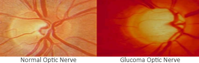
Glaucoma is a disease that damages the optic nerve. The optic nerve carries electrical impulses of the light images transformed by the retina, to our brain. Damage to the optic nerve and retina causes blind spots in the field of vision. In extreme cases, if the entire nerve is destroyed, blindness will occur. Glaucoma is in fact one of the leading causes of blindness and affects an estimated one of every 50 adults.

Although glaucoma can occur at any age, the risk of developing this increases dramatically after the age of 35.

Image frame 2

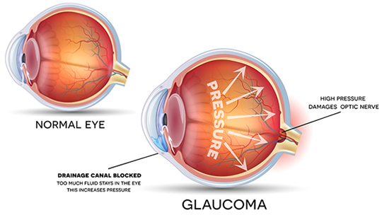
High Pressure on Optic Nerves

At Khandwala's Eyecare, We will check your IOP and evaluate your optic nerve. If your eye pressure is elevated or your optic nerve looks suspicious, We will likely perform visual field tests and specialized
scans of your retina and optic nerve to determine if you have glaucoma.

Symptoms of Glaucoma..
At first, open-angle glaucoma has no symptoms. It causes no pain. Vision stays normal. Glaucoma can develop
in one or both eyes. Without treatment,people with glaucoma will slowly lose their peripheral (side) vision. As glaucoma remains untreated, people may miss objects to the side and out of the corner of their eye. They seem to be looking through a tunnel. Over time, straight-ahead (central) vision may decrease until no vision remains.


A Walkthough Video of Glaucoma..
Glaucoma Risk Increases in Families: Spread the Word..

Glaucoma is a worldwide problem that can lead to blindness. It is especially problematic because there are often no symptoms in its early stages. It is estimated that up to 50 percent of people with glaucoma don't realize they have it.Numerous population-based studies have demonstrated that one of the greatest risk factors for glaucoma is a family history of the disease. That means that one of the most important things you can do is to talk about glaucoma with your family and encourage them to take steps to preserve their vision.People at high risk for glaucoma should get a complete eye exam, including eye dilation, every one or two years.Glaucoma is much more common among older people. You are six times more likely to get glaucoma if you are over 60 years old.

Types of Glaucoma..

The two major categories of glaucoma are Open-angle glaucoma (OAG) and Narrow angle glaucoma (NOG).
The "angle" in both cases refers to the drainage angle inside the eye that controls the outflow of the watery fluid (aqueous) that is continually being produced inside the eye.
If the aqueous can access the drainage angle,the glaucoma is known as open angle glaucoma. If the drainage angle is blocked and the aqueous cannot reach it, the
glaucoma is known as narrow angle glaucoma.
Variations of OAG include: primary open angle glaucoma (POAG), normal-tension glaucoma (NTG), pigmentary glaucoma, pseudoexfoliation glaucoma, secondary glaucoma
and congenital glaucoma.Variations of narrow angle glaucoma include include acute angle closure glaucoma, chronic angle closure glaucoma, and neovascular glaucoma.

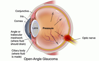
  1. The most common form of glaucoma, accounting for
    at least 90% of all glaucoma cases:
  2. Is caused by the slow clogging of the drainage canals, resulting in increased eye pressure.
  3. Has a wide and open angle between the iris and cornea.
  4. Develops slowly and is a lifelong condition.
  5. Has symptoms and damage that are not noticed.
  6. Family history of glaucoma.
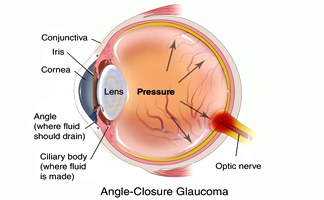
  1. Angle-closure glaucoma, a less common form of glaucoma:
  2. Is caused by blocked drainage canals, resulting in a sudden rise in intraocular pressure.
  3. Has a closed or narrow angle between the iris and cornea.
  4. Has symptoms and damage that are usually very noticeable.
  5. Demands immediate medical attention.
  6. Develops very quickly.

More Categories :

Are there other types of Glaucoma ?

Yes.

Although there is only two major types of Glaucoma : Open Angle and Narrow Angle : futher below are the some more categories related to both.
Primary open-angle glaucoma.
This common type of glaucoma gradually reduces your peripheral vision without other symptoms. By the time you notice it, permanent damage already has occurred.If your IOP remains high, the destruction caused by POAG can progress until tunnel vision develops, and you will be able to see only objects that are straight ahead. Ultimately, all vision can be lost, causing blindness.
Acute angle-closure glaucoma.
Also called narrow-angle glaucoma, acute angle-closure glaucoma produces sudden symptoms such as eye pain, headaches, halos around lights, dilated pupils, vision loss, red eyes, nausea and vomiting.These signs constitute a medical emergency. The attack may last for a few hours, and then return again for another round, or it may be continuous without relief. Each attack can cause progressively more vision loss.
Normal-tension glaucoma.
Like POAG, normal-tension glaucoma (also called normal-pressure glaucoma, low-tension glaucoma or low-pressure glaucoma) is a type of open-angle glaucoma that can cause visual field loss due to optic nerve damage. But in normal-tension glaucoma, the eye's IOP remains in the normal range.Also, pain is unlikely and permanent damage to the eye's optic nerve may not be noticed until symptoms such as tunnel vision occur.The cause of normal-tension glaucoma is not known. But many doctors believe it is related to poor blood flow to the optic nerve. Normal-tension glaucoma is more common in those who are Japanese, are female and/or have a history of vascular disease.
Pigmentary glaucoma.
This rare form of glaucoma is caused by clogging of the drainage angle of the eye by pigment that has broken loose from the iris, reducing the rate of aqueous outflow from the eye. Over time, an inflammatory response to the blocked angle damages the drainage system.You are unlikely to notice any symptoms with pigmentary glaucoma, though some pain and blurry vision may occur after exercise. Pigmentary glaucoma most frequently affects white males in their mid-30s to mid-40s.
Secondary glaucoma.
Symptoms of chronic glaucoma following an eye injury could indicate secondary glaucoma, which also may develop with presence of eye infection, inflammation, a tumor or enlargement of the lens due to a cataract.
Congenital glaucoma.
This inherited form of glaucoma is present at birth, with 80 percent of cases diagnosed by age one. These children are born with narrow angles or some other defect in the drainage system of the eye.It's difficult to spot signs of congenital glaucoma, because children are too young to understand what is happening to them. If you notice a cloudy, white, hazy, enlarged or protruding eye in your child, consult your eye doctor. Congenital glaucoma typically occurs more in boys than in girls.
How is Glaucoma Detected ?

Glaucoma is detected through a comprehensive dilated eye exam that includes the following:

Visual acuity test : An Eye chart test measures how well you see at various distances.

Visual field test: This test measures your peripheral (side vision). It helps your eye care professional
tell if you have lost peripheral vision, a sign of glaucoma.

Dilated eye exam : In this exam, drops are placed in your eyes to widen, or dilate, the pupils. Your eye
care professional uses a special magnifying lens to examine your retina and optic nerve for signs of
damage and other eye problems. After the exam, your close-up vision may remain blurred
for several hours.

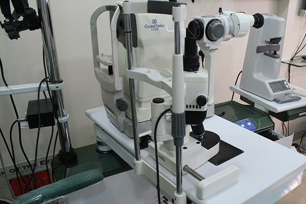
Tonometry : Tonometry is the measurement of pressure inside the eye by using an instrument called a tonometer. Numbing drops may be
applied to your eye for this test. A tonometer measures pressure inside the eye to detect glaucoma.

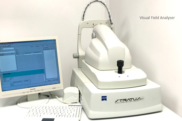
Oct : Screening with Ocular Coherence Tomography (OCT) enables diagnoses of potentially serious conditions that can affect your eyesight.
An advanced eye scan, OCT, uses light to illustrate the different layers that make up the back of your eye. In OCT scan, a digital photograph
and a three dimensional cross sectional scan of the back of the eye are obtained. At Khandwala Eyecare the OCT scan is used extensively in
detecting if you are at risk of glaucoma or the stage of glaucoma you are at. OCT is also used to detect, identify and monitor the progress of
age-related macular degeneration and macular hole. It also enables the early detection and treatment of diabetic retinopathy and
vitreomacular traction.

Pachymetry : is the measurement of the thickness of your cornea. Your eye care professional applies a numbing drop to your eye and uses an ultrasonic wave instrument to measure the thickness of your cornea.

Glaucoma Treatments..

Immediate treatment for early-stage, open-angle glaucoma can delay progression of the disease. That’s why early diagnosis is very important.
Glaucoma treatments include :Medicines, Laser Trabeculoplasty, Conventional Surgeryor a combination of any of these. While these treatments may save remaining vision, they do not improve sight already lost from glaucoma.

Medicines : Medicines, in the form of eyedrops or pills, are the most common early treatment for glaucoma. Taken regularly,these eyedrops lower eye pressure. Some medicines cause the eye to make less fluid. Others lower pressure by helping fluid drain from the eye. Before you begin glaucoma treatment,tell your eye care professional about other medicines and supplements that you are taking. Sometimes the drops can interfere
with the way other medicines work. Glaucoma medicines need to be taken regularly as directed by your eye
care professional. Most people have no problems, However,some medicines can cause headaches or
other side effects. For example, drops may cause stinging, burning, and redness in the eyes.
Many medicines are available to treat glaucoma.If you have problems with one medicine, tell your eye care professional. Treatment with a different
dose or a new medicine may be possible. Because glaucoma often has no symptoms, people may be tempted to stop taking, or may forget to take, their medicine. You need to use the drops or pills as long as they help control your eye pressure. Regular use is very important. Make sure your eye
care professional shows you how to put the drops into your eye. For tips on using your glaucoma eyedrops, see the inside or back cover of its booklet.

Laser Trabeculoplasty : Laser trabeculoplasty helps fluid drain out of the eye. Your doctor may suggest this step at any time. In many cases, you will need to keep taking glaucoma medicines after this procedure.Laser trabeculoplasty is performed in your doctor’s office or eye clinic. Before the surgery, numbing drops are applied to your eye. As you sit facing the laser machine, your doctor holds a special lens to your eye. A high-intensity beam of light
is aimed through the lens and reflected onto the meshwork inside your eye. You may see flashes of bright green or red light. The laser makes several evenly spaced burns that stretch the drainage holes in the meshwork. This allows the fluid to drain better.Like any surgery, laser surgery can cause side effects, such as inflammation. Your doctor may give you some drops to take home for any soreness or inflammation inside the eye. You will need to make several follow-up visits to have your eye pressure and eye monitored.If you have glaucoma in both eyes, usually only one eye will be treated at a time. Laser treatments for each eye will be scheduled several days to several weeks apart.Studies show that laser surgery can be very good at reducing the pressure in some patients. However, its effects can wear off over time. Your doctor may suggest further treatment.

Conventional Surgery : Conventional surgery makes a new opening for the fluid to leave the eye. Your doctor may suggest this treatment at any time. Conventional surgery often is done after medicines and laser surgery have failed to control pressure.Conventional surgery, called trabeculectomy, is performed in an operating room. Before the surgery, you are given medicine to help you relax. Your doctor makes small injections around the eye to numb it. A small piece of tissue is removed to create a new channel for the fluid to drain from the eye. This fluid will drain between the eye tissue
layers and create a blister-like “filtration bleb.”For several weeks after the surgery, you must put drops in the eye to fight infection and inflammation. These drops will be different from those you may have been using before surgery. Conventional surgery is performed on one eye at a time.

Usually the operations are four to six weeks apart Conventional surgery is about 60 to 80 percent effective at lowering eye pressure. If the new drainage opening narrows, a second operation may be needed. Conventional surgery works best if you have not had previous eye surgery,such as a cataract operation. act, problems with the cornea, inflammation, infection inside the eye, or low eye pressure problems. If you have any of these problems, tell your doctor so a treatment plan can be developed.